Biology · Kidney
The juxtaglomerular apparatus: three cells, two jobs
In every nephron the tubule loops back and touches the arteriole that feeds its own glomerulus. That contact point is a sensor, and it helps set your blood pressure. The video tours it on a real kidney section; the notes below sort out which cell does what, and the board is yours to copy.
By Boardling, from the Instaboard teamLesson 005 · 3:03 videoUpdated
- 0:00Why does the tubule touch its own blood vessel?
- 0:21A real kidney section: where the JGA is
- 0:46The three cells
- 0:49Macula densa
- 1:00JG cells and renin
- 1:16Extraglomerular mesangial cells
- 1:26Job 1: protecting the filter
- 1:38Job 2: blood pressure
- 1:54Common mix-up: where renin comes from
- 2:02Your turn
- 2:21Back to the question
Juxtaglomerular apparatus in 30 seconds
- The juxtaglomerular apparatus (JGA) is where the distal tubule touches the afferent arteriole of its own glomerulus. "Juxta" means next to.
- Macula densa: packed cells in the distal tubule wall that sense the salt (NaCl) and flow in the filtrate.
- JG cells: modified smooth muscle in the afferent arteriole wall that sense pressure and store and release renin.
- Extraglomerular mesangial cells sit in the gap between the two and relay signals.
- Two jobs: too much salt arriving → the afferent arteriole narrows and GFR falls; low pressure or low salt → renin → angiotensin II → blood pressure up.
The notes board from the lesson
This is the board built during the video. Copy it and it's yours: add your own cards, rearrange it, draw on it.
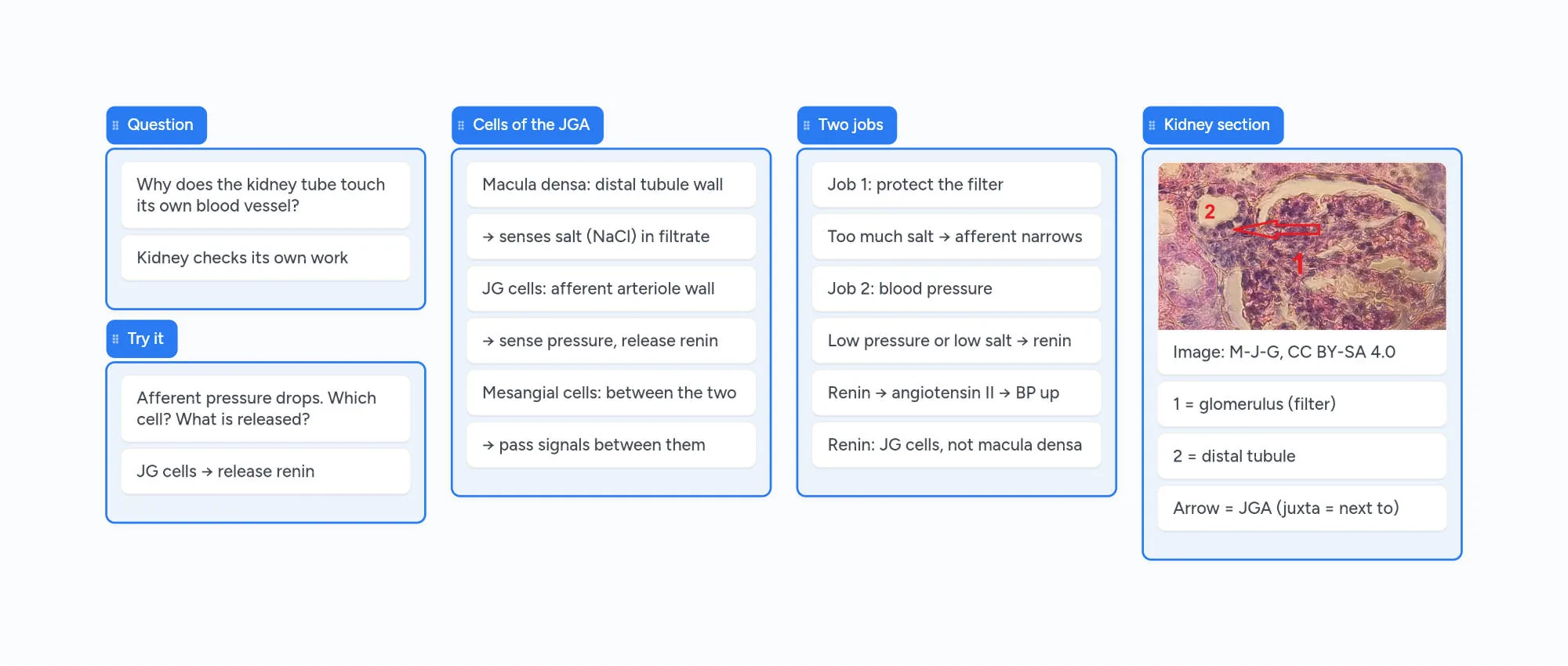
Why does the tubule touch its own blood vessel?
In the video at 0:00
Follow one nephron and something looks odd. After the loop of Henle, the tubule climbs back up and presses against the arteriole that brings blood into its own glomerulus. It isn't an accident of packing. That contact point is a sensor the kidney uses to check its own work.
The structure at that spot is the juxtaglomerular apparatus, and it is built from just three kinds of cells.
Finding it on a real kidney section
In the video at 0:21
The micrograph on the board is a stained slice of a pig's kidney, which is organised the same way as ours. Label 1 is a glomerulus, the knot of capillaries that filters the blood. Label 2 is the distal tubule, carrying filtrate back up. The arrow marks where the two touch: the juxtaglomerular apparatus.
The three cells and where each one sits
In the video at 0:46
Macula densa. A patch of tightly packed cells in the wall of the distal tubule, right where it meets the arterioles. They monitor the sodium chloride in the filtrate flowing past, and each has a single cilium that bends with the flow rate. They don't make renin; they sense and send signals.
Juxtaglomerular (JG) cells. Modified smooth muscle cells in the wall of the afferent arteriole, the vessel feeding the glomerulus. They feel the pressure in that vessel directly, can contract to narrow it, and store the enzyme renin in granules (which is why they're also called granular cells).
Extraglomerular mesangial cells. They fill the small space between the macula densa and the arterioles and are thought to carry signals between the other two cell types.
Job 1: protecting the filter
In the video at 1:26
If the glomerulus filters too fast, filtrate rushes through the tubule with less time for salt to be taken back, so more NaCl reaches the macula densa. The macula densa answers with a local signal that makes the afferent arteriole narrow. Less blood enters the glomerulus and the filtration rate (GFR) drops back down. This loop is called tubuloglomerular feedback.
Job 2: blood pressure through renin
In the video at 1:38
When pressure in the afferent arteriole falls, or the macula densa detects too little salt, the JG cells release renin into the blood. Renin turns angiotensinogen into angiotensin I, which is then converted to angiotensin II. Angiotensin II narrows blood vessels and raises blood pressure, and it also triggers aldosterone so more sodium and water are kept.
So a small sensor, repeated in every nephron, feeds into one of the body's main blood-pressure systems.
Practice question
The macula densa detects far more NaCl than normal arriving in the distal tubule. Does the afferent arteriole widen or narrow, and what happens to GFR?
Show the answer
It narrows, so GFR falls. More salt arriving means the glomerulus is filtering too fast. Narrowing the afferent arteriole lets less blood into the glomerulus, so less is filtered.
Common mix-ups
The macula densa releases renin.
Renin comes from the JG cells in the afferent arteriole wall. The macula densa only senses salt and signals.
The JGA is part of the glomerulus.
It sits next to the glomerulus, at the point where the distal tubule meets the afferent arteriole.
JG cells sit in the efferent arteriole.
They line the afferent arteriole, the vessel bringing blood into the glomerulus, so they feel its pressure first.
How this lesson was made
Boardling lessons are made by the Instaboard team. The narration is a synthetic (text-to-speech) voice, and the script and these notes were written with AI help, then checked against the sources listed below (last checked October 30, 2026). The notes board is built live in Instaboard during the video. Spotted a mistake? Tell us and we'll fix it.
Put this lesson in your notes
The board opens in your browser in a few seconds. Add your lecture notes, photos of your handwritten pages and your own questions next to it. Sign in for free to keep it.
Questions
What does the juxtaglomerular apparatus do?
It does two jobs. It keeps the filtration rate of its own nephron steady by narrowing the afferent arteriole when too much salt reaches the macula densa (tubuloglomerular feedback). And when blood pressure or salt delivery falls, its JG cells release renin, which leads to angiotensin II and higher blood pressure.
What are the three cells of the juxtaglomerular apparatus?
The macula densa cells in the wall of the distal tubule, the juxtaglomerular (granular) cells in the wall of the afferent arteriole, and the extraglomerular mesangial cells in the space between them.
What is the difference between the macula densa and JG cells?
Macula densa cells are tubule cells that sense the sodium chloride in the filtrate. JG cells are modified smooth muscle cells in the afferent arteriole that sense blood pressure and release renin.
What triggers renin release?
A drop in pressure in the afferent arteriole, a fall in the salt reaching the macula densa, and sympathetic nerve signals during low blood pressure all make the JG cells release renin.
Where is the juxtaglomerular apparatus located?
At the vascular pole of each renal corpuscle, where the first part of the distal convoluted tubule comes back and touches the afferent (and efferent) arteriole of its own glomerulus.
Exam fit
- MCAT 3B: excretory system
- Anatomy & physiology
- Renal physiology