Biology · Muscle and nerve physiology
Sliding filament theory: how muscles contract, step by step
Bend your arm and the muscle gets shorter, yet the threads inside it never do. The video builds the answer live on a board: the two kinds of threads, the hooks that grab and pull, why ATP is needed to let go, and how calcium switches the whole thing on. The notes below add the textbook terms, and the board is yours to copy.
By Boardling, from the Instaboard teamLesson 012 · 2:19 videoUpdated
Sliding filament theory in 30 seconds
- Each muscle fiber is built from repeating sections called sarcomeres, each with thin threads (actin) and thick threads (myosin).
- The myosin heads grab the actin (a cross-bridge) and pull it toward the middle of the sarcomere. The threads slide past each other; neither thread gets shorter, but the sarcomere does.
- A fresh ATP makes the myosin head let go, and breaking that ATP down re-cocks the head for the next pull. With no ATP the heads stay stuck, which is why muscles go stiff after death (rigor mortis).
- Calcium is the switch: it binds to troponin, so tropomyosin slides off the binding spots on actin and the heads can grab. When the nerve signal stops, calcium is pumped back and the muscle relaxes.
The notes board from the lesson
This is the board built during the video. Copy it and it's yours: add your own cards, rearrange it, draw on it.
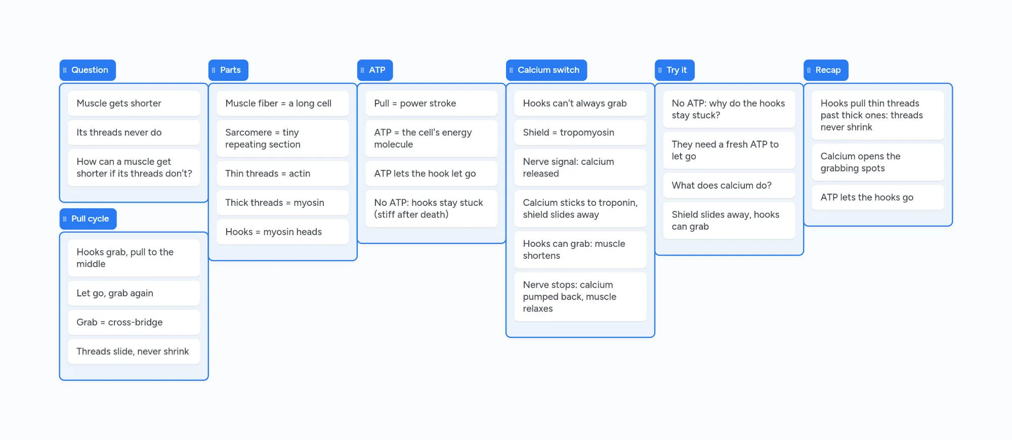
The muscle shortens, its threads don't
In the video at 0:00
Bend your arm and the muscle in it gets shorter. Yet the protein threads inside it keep the same length the whole time. The answer to that puzzle is the sliding filament model of muscle contraction.
Fibers, sarcomeres, actin and myosin
In the video at 0:14
A skeletal muscle is made of long cells called muscle fibers. Inside each fiber are long strands called myofibrils, and each myofibril is a chain of tiny repeating sections called sarcomeres, the units that actually shorten.
Every sarcomere has two kinds of threads (filaments): thin actin filaments anchored at the ends of the sarcomere (the Z-discs), and thick myosin filaments anchored in the middle (the M-line). The thick filaments have myosin heads sticking out, the hooks in the video.
Hooks grab and pull: the cross-bridge
In the video at 0:33
A myosin head binds to a spot on the actin; that link is a cross-bridge. The head pulls the thin filament toward the center of the sarcomere, lets go, and grabs again further along. OpenStax compares it to rowing: the oars pull, lift out of the water, reset and pull again. This repeated loop is the cross-bridge cycle.
The filaments slide past each other, so the Z-discs move closer together and the sarcomere shortens. All the sarcomeres in a fiber shorten together, so the whole fiber and the muscle shorten. In exam terms, the I band gets smaller while the A band stays the same width.
The power stroke and ATP
In the video at 0:55
The pull itself is the power stroke: the head pivots toward the middle and moves the thin filament about 10 nanometres. Each head can only pull that short way before it has to be reset.
To let go, the head needs a fresh molecule of ATP, the cell's energy molecule. ATP binding makes the head detach from actin; breaking the ATP down into ADP and phosphate re-cocks the head, ready for the next pull.
With no ATP, the heads cannot detach and the cross-bridges stay in place. That is the cause of rigor mortis, the stiffness of skeletal muscles soon after death.
Calcium: the switch
In the video at 1:15
The heads can't grab at any time. At rest a protein called tropomyosin winds along the actin and covers the binding spots, held in place with another protein, troponin.
A motor neuron signal (the neurotransmitter acetylcholine) starts an electrical signal that spreads along the fiber's membrane and into its T-tubules. That makes the sarcoplasmic reticulum, the fiber's calcium store, release calcium. Calcium binds to troponin, tropomyosin slides off the binding spots, and the heads can grab.
When the nerve signal stops, pumps powered by ATP move calcium back into the sarcoplasmic reticulum, tropomyosin covers the spots again, and the fiber relaxes.
Back to the question
In the video at 2:01
A muscle gets shorter because myosin heads pull the thin actin filaments past the thick myosin filaments, so every sarcomere shortens while the filaments keep their length. Calcium opens the binding spots, and ATP lets the heads let go so they can pull again.
How the force of a whole muscle is graded is covered in the lessons on twitch, summation and tetanus and motor unit recruitment.
Practice question
A muscle cell runs out of ATP. Why do the myosin heads stay stuck to actin?
Show the answer
A myosin head needs a fresh ATP to let go of actin. Without ATP the cross-bridges can't break, so the heads stay attached. ATP binding is the step that detaches the head from actin, and breaking ATP down re-cocks it. With no ATP the cycle stops with the heads attached, which is what happens in rigor mortis.
Common mix-ups
During contraction the actin and myosin filaments get shorter.
Neither filament changes length. The thin filaments slide past the thick ones, so the sarcomere shortens while the filaments stay the same.
ATP is used to make the myosin head grab actin.
ATP binding makes the head let go of actin. Breaking that ATP down re-cocks the head for the next pull.
Calcium binds to tropomyosin to uncover the binding sites.
Calcium binds to troponin. That moves tropomyosin away from the binding sites on actin.
How this lesson was made
Boardling lessons are made by the Instaboard team. The narration is a synthetic (text-to-speech) voice, and the script and these notes were written with AI help, then checked against the sources listed below (last checked November 9, 2026). The notes board is built live in Instaboard during the video. Spotted a mistake? Tell us and we'll fix it.
Put this lesson in your notes
The board opens in your browser in a few seconds. Add your lecture notes, photos of your handwritten pages and your own questions next to it. Sign in for free to keep it.
Questions
What is the sliding filament theory?
The model of muscle contraction in which myosin heads pull the thin actin filaments past the thick myosin filaments, shortening each sarcomere while the filaments themselves keep their length.
What is a cross-bridge?
The link formed when a myosin head binds to a binding site on an actin filament. Cross-bridges form and break over and over during contraction.
What is the power stroke?
The step in which the attached myosin head pivots toward the center of the sarcomere and pulls the thin filament about 10 nm.
What does calcium do in muscle contraction?
Calcium released from the sarcoplasmic reticulum binds to troponin, which moves tropomyosin off the binding sites on actin so myosin heads can attach.
Why does rigor mortis happen?
After death no new ATP is made. Myosin heads need ATP to detach from actin, so the cross-bridges stay in place and the muscles go stiff.
Exam fit
- A&P
- AP Biology
- MCAT biology
Sources
- OpenStax Anatomy & Physiology 2e, 10.3 Muscle Fiber Contraction and Relaxation: sliding filament model, cross-bridge cycle, ATP, calcium, troponin and tropomyosin, rigor mortis (checked November 9, 2026)
- OpenStax Anatomy & Physiology 2e, 10.2 Skeletal Muscle: muscle fibers, myofibrils, sarcomeres, sarcoplasmic reticulum and T-tubules (checked November 9, 2026)
More on the muscle and nerve physiology
Twitch, summation and tetanus
How to read a myogram: one twitch, wave summation, unfused and fused tetanus, and why calcium makes twitches add up.
Motor unit recruitment
How a muscle grades its force: motor units, small versus large units, the size principle, and the two ways to turn force up.
Action potential
Every step of the action potential graph for the MCAT: resting potential, threshold, depolarization, the +30 mV peak, repolarization, hyperpolarization and the refractory period.